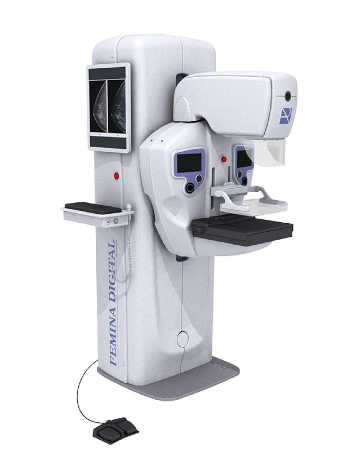

FEMINA DIGITAL
DIGITAL MAMMOGRAPHY DEVICE
IMPACT WITH AN EARLY DIAGNOSIS
FEMINA DİJİTAL, has been designed to meet all diagnostic requests, with different configurations allowing the radiologist to use the latest digital mammography imaging methods.The designed servo assisted isocentric C-arm makes the system more efficient and superior in terms of diagnostic efficiency.
FEMINA DİJİTAL, relies on a specialized platform that facilitates all the necessary tools that optimize the process for screening and diagnostic processes. A functional and ergonomic design provides more comfortable patient positioning.This is complemented by a fast, user friendly operating system that completes the process and reduces the discomfort that such examinations can cause.
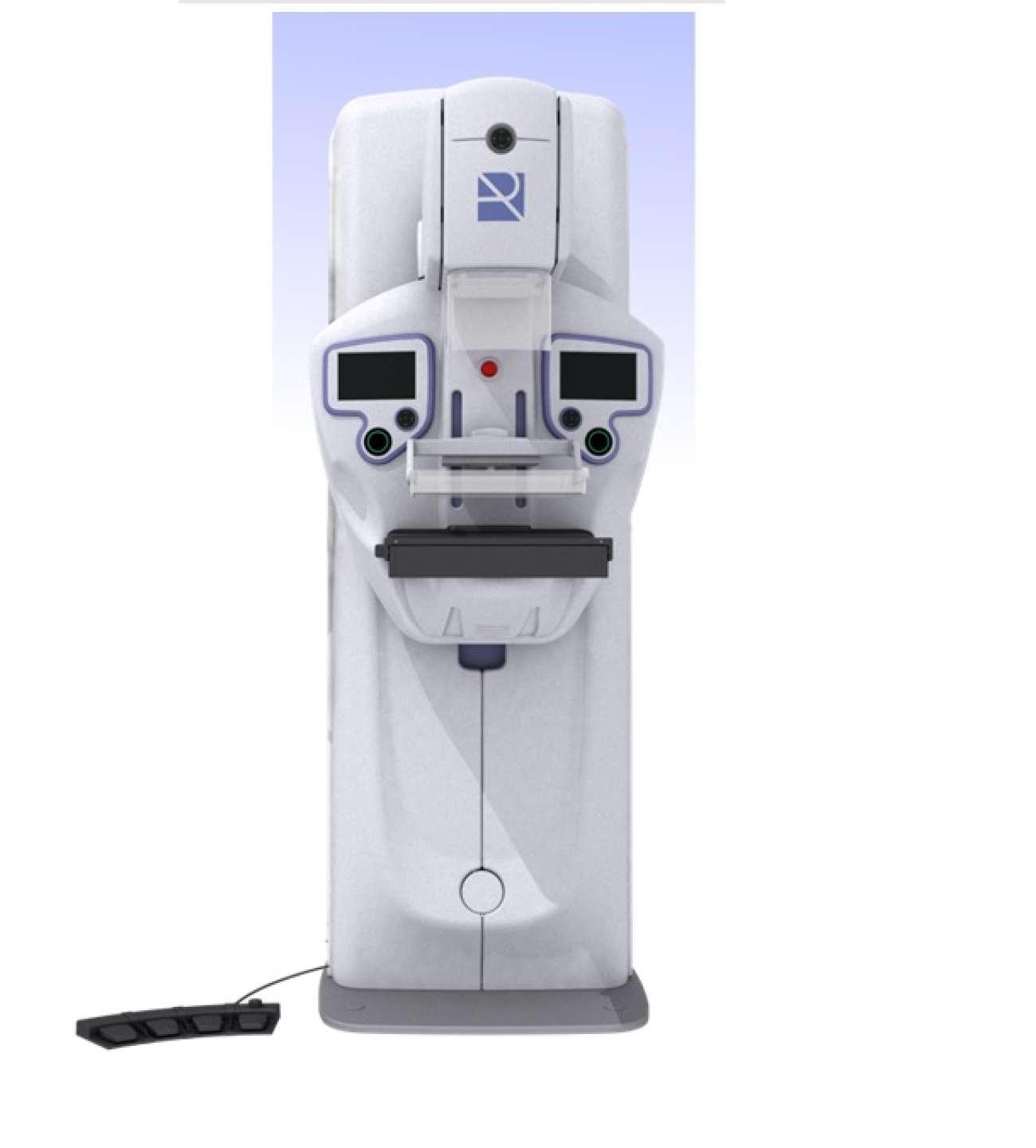
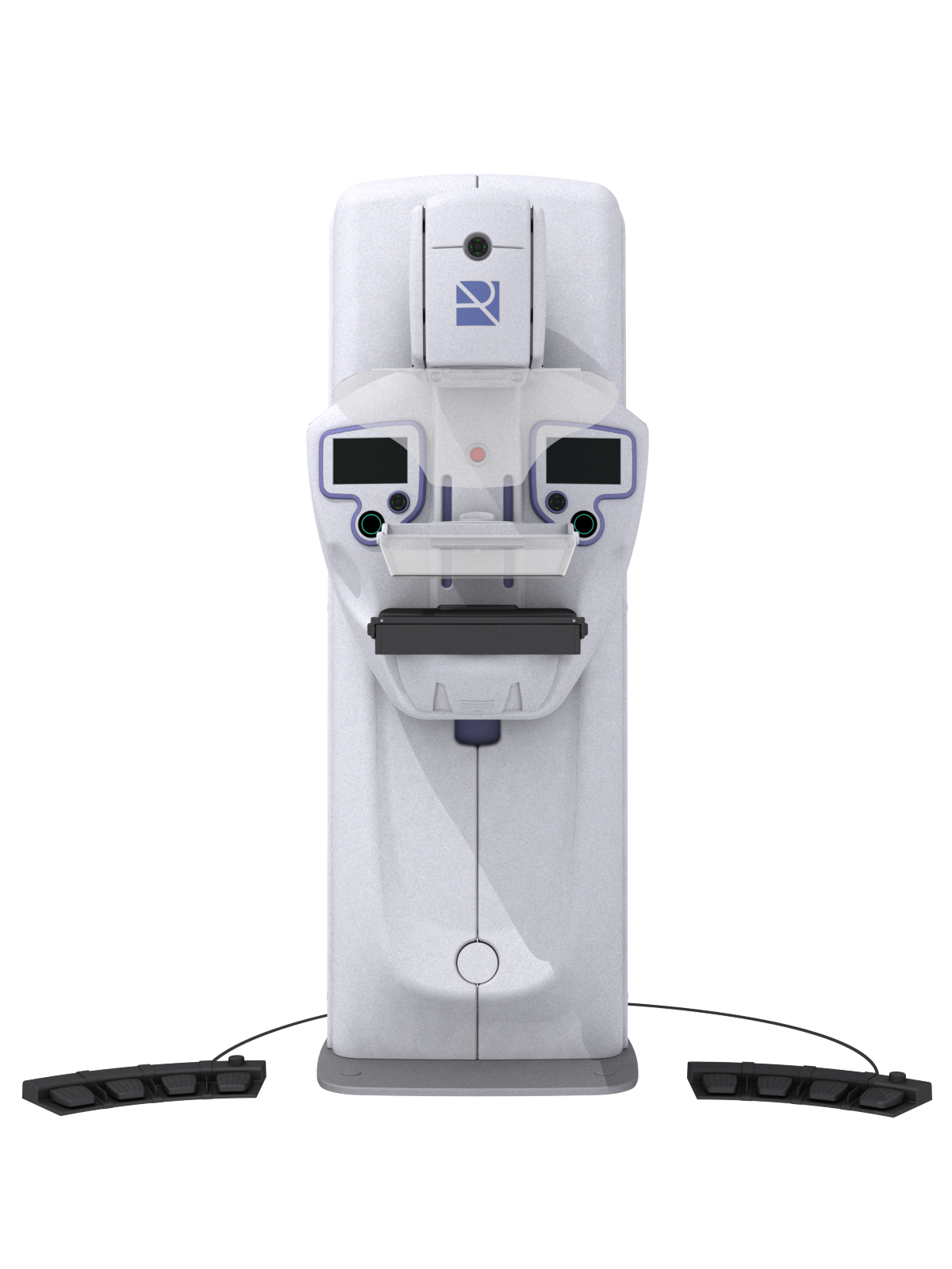
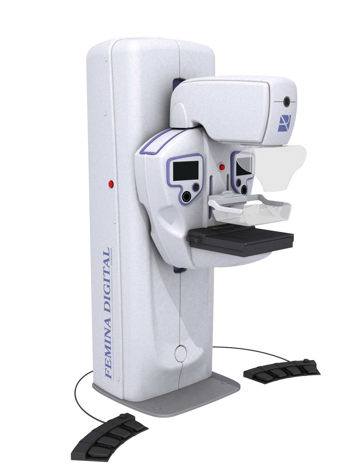
Isocentric C Arm: For breast projections and height adjustment without moving the patient .
"Smart mpress" Compression System: Ensures an optimal compression of the breast with minimal discomfort for the patient. Special "FTSE" function automaticly adjusts the force to be applied according to breast density.
"Sensoroi" Automatic Exposure Control: Dual operating mode which sets the exposure parameters based on breast density and breast thickness.
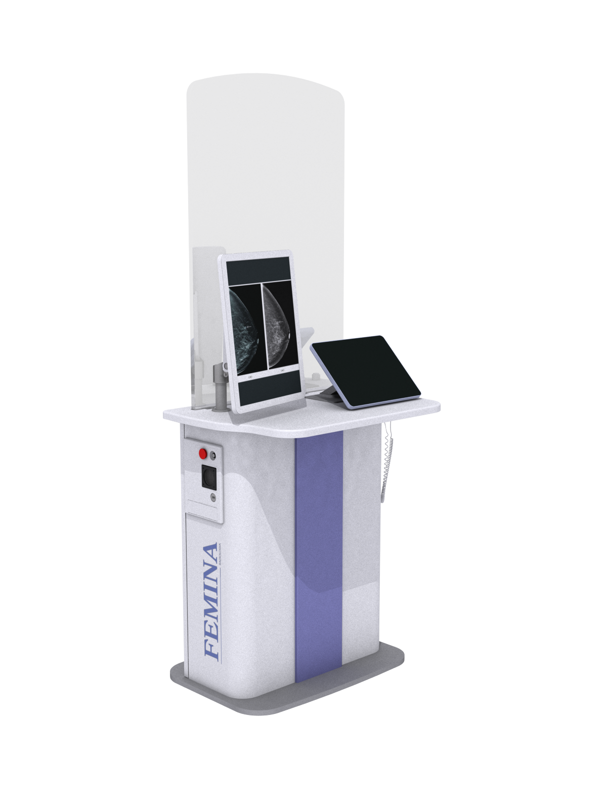
Optimized Workflow: Optional remote acqusition workstation with anti-x protection barrier with 15" touch screen colour display and 24" TFT LCD color monitor for image visualization.
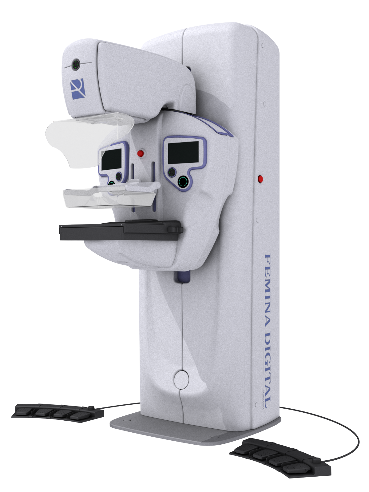
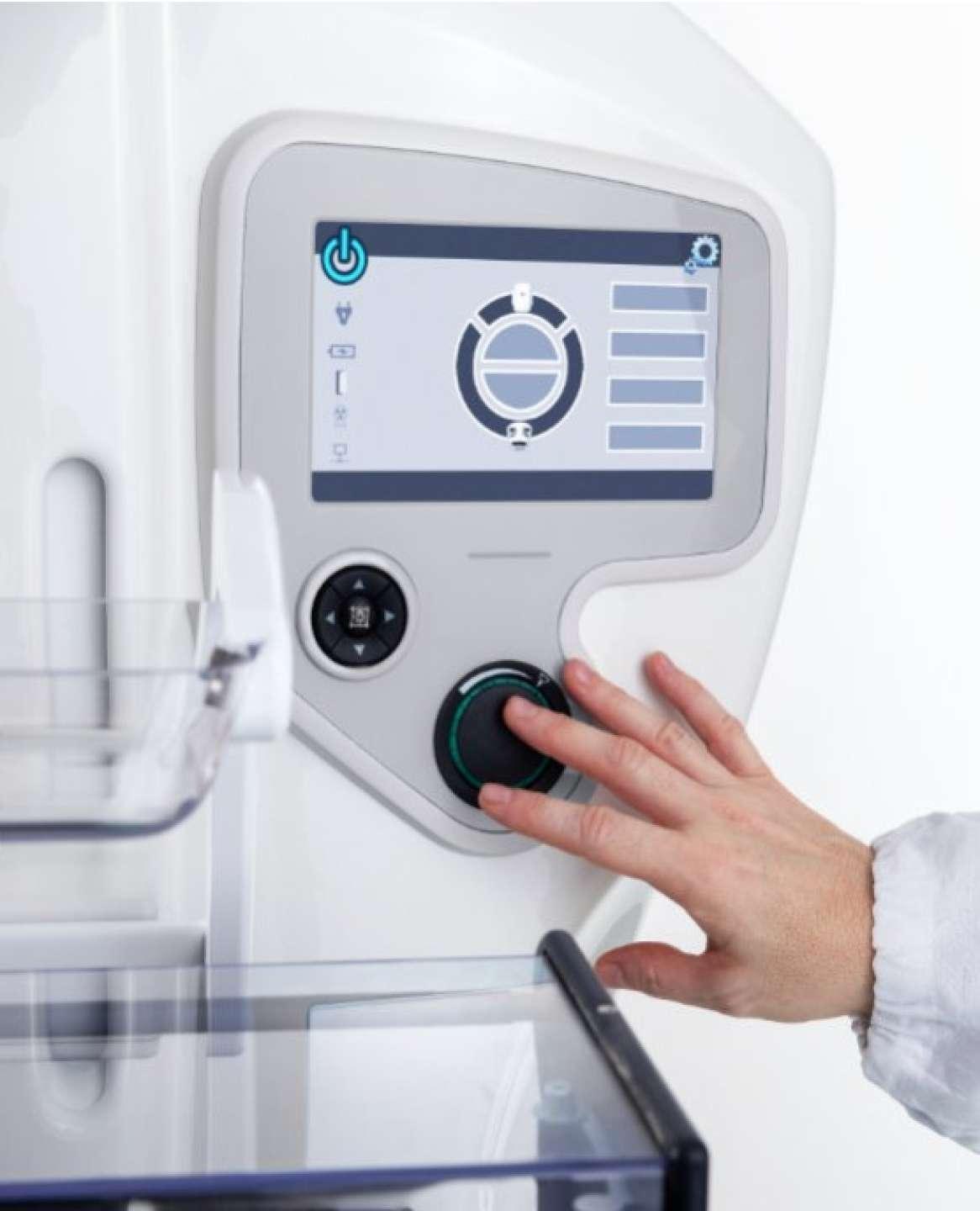
7" Touch Screen Colour Displays : Two colour touch screen displays located on both side of the C-arm to show informations such as compression force, compressed breast thickness, collimation format, magnification factor, projection angle, patient name.
Pedal Footswitch: Foot pedal controls for fine tuning compression and release of compression.
There are Two colour touch screen displays located on both side of the C-arm to show informations such as compression force, compressed breast thickness, collimation format, magnification factor, projection angle, patient name.Operator can select breast laterality and anatomical programs through these screens.
Four multiple switches for an optimal workflow assists vertical and rotational motion control through continuous, pre-programmed selections.
In the centre is placed the collimator light (after 20 seconds automaticly switched off).
Two rotary controllers allow the operator to perform a manual fine adjustment for additional adjustments after compression.
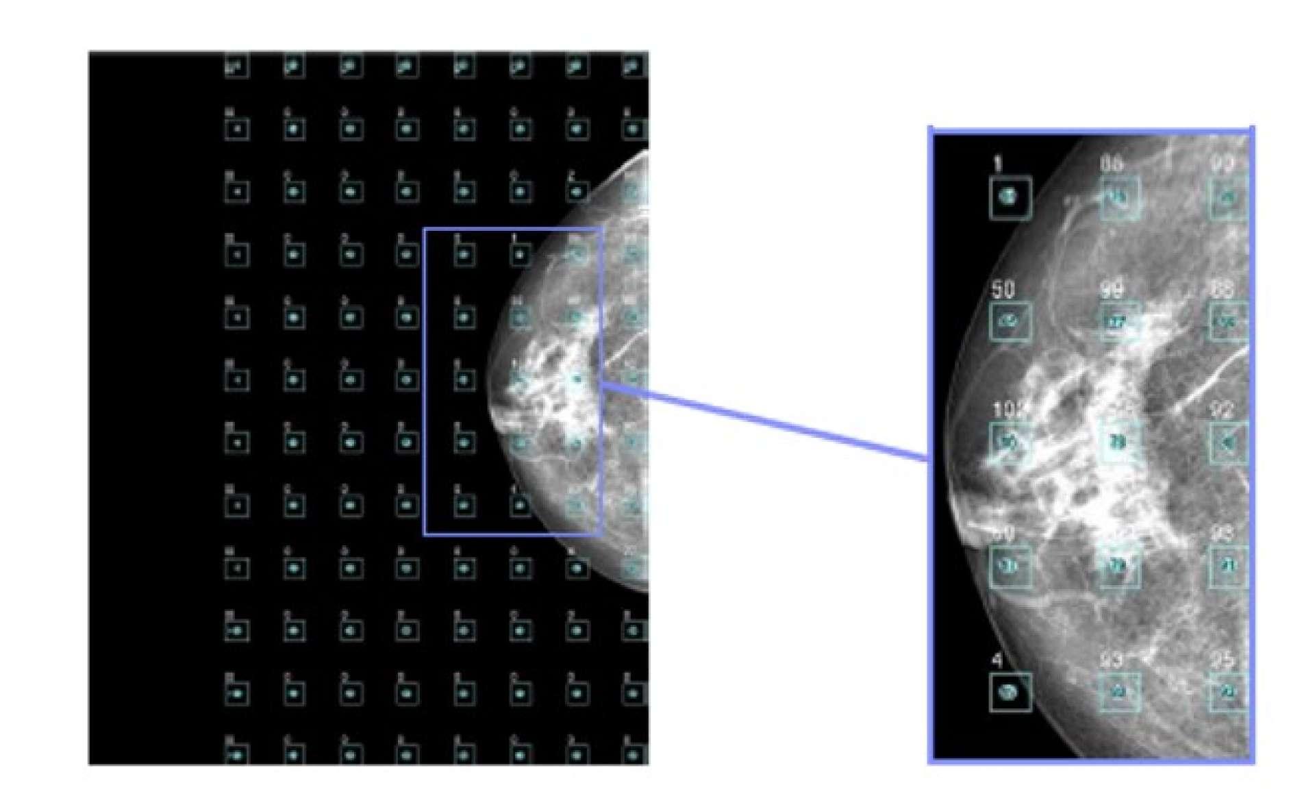
Automatic exposure system "SENSOROI", guarrantees the optimal selection of the exposure technique for any condition, including special cases with breast implant or presense of metal surgical elements.
The AEC system executes a very short and very low dose pre-exposure and the digital system software analyses the pre-image obtained, based on the analysis of 96 different areas selects the actual exposure technique that is executed next.This method ensures that it is taking the composition and density of the breast and not only its size
In addition, a fast working mode, can also be selected, choosing an exposure technique based on the thickness of the compressed breast, this mode allows the study to be carried out in less time when it is necessary to keep the compression as short as possible.The advanced calculation algorithm provides optimal results in all breast types and conditions.
Smart Compression System
Motorized compression system controlled by microprocessor.Buit in sensors detect breast density and automaticly adapt the compression force.Additionaly, the compression speed decreases proportionally as the breast is compressed.
The equipment, can be configured for a maximum compression force of 15kg or 20kg according to the requirements.It includes a triple safety system to avoid excessive compression. The compression paddle, is automaticly released after each exposure, the operator can inhibit this mode of operation by selecting it on the control screen for example, 2D biopsy procedures.
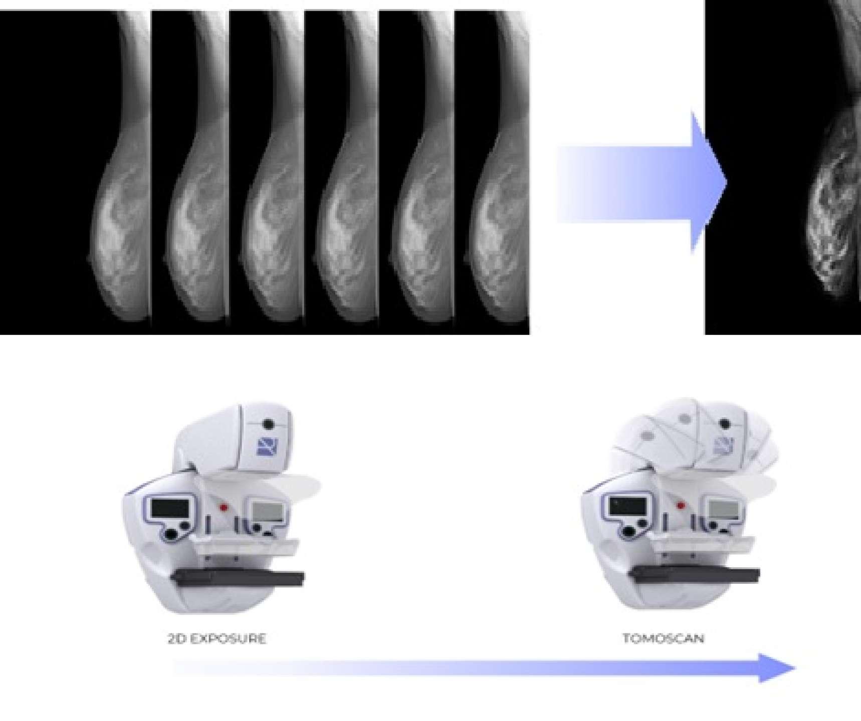
While scanning the breast, the system takes a series of low dose x-ray exposures at different angles. Through sophisticated algorithm, allows volumetric breast tissue reconstruction(3D).
The outstanding image quality, allows a clear display of lesions and areas of interest and offers radiologists the best diagnostic accuracy with a low level of radiation as compared to convanctional mammography .
Software produces 2D images directly from tomosynthesis images, without administering an additional dose to patient.
IMAGE ACQUSITION WORKSTATION
Acqusition workstation equipped with transparent x-ray protection screen to stand next to the patient during acqusition while providing the working area X-Ray protection.
*0,34mm Pb Anti-X protection barrier.
*24" 2MP color monitor
*15" colour control panel.
*CD/DVD/USB/Speaker
*7,4kW Generator
*Tungsten X-Ray Tube 0,1/0,3
*Motorised Isocentric Carm
*Biangular anode
*Automatic Collimator.
*24x30 cm Silicium or Selenium detector
*2 7" TFT Led colour touch screen
*Removable Grid
* Multipurpose foot pedal

Download Brochure